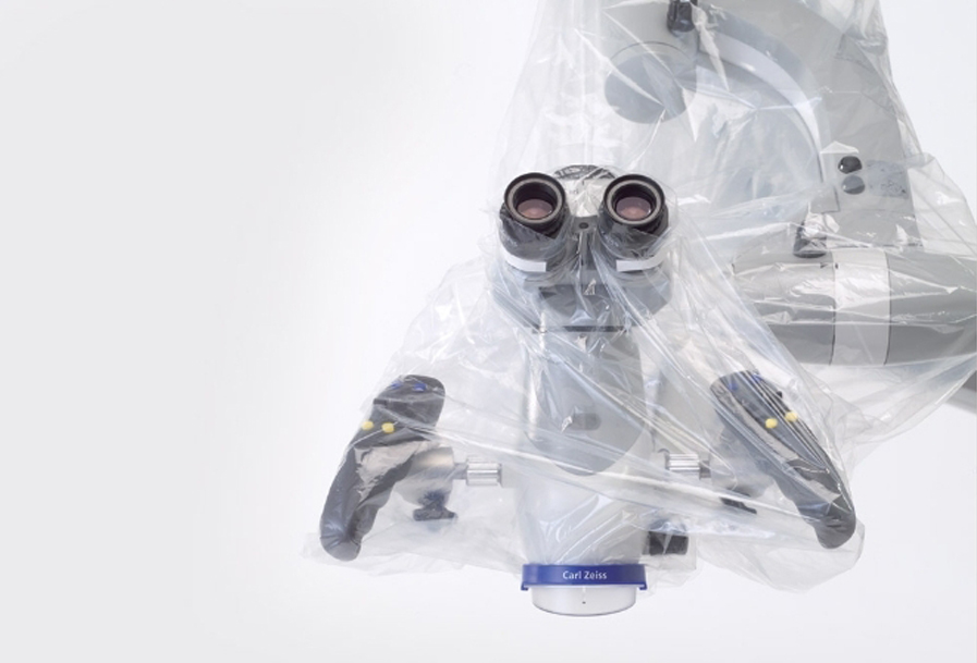
The Microscope
Doing something in the mouth is a big issue. Everything is very small.
No light, no room.
Only the microscope helps you:
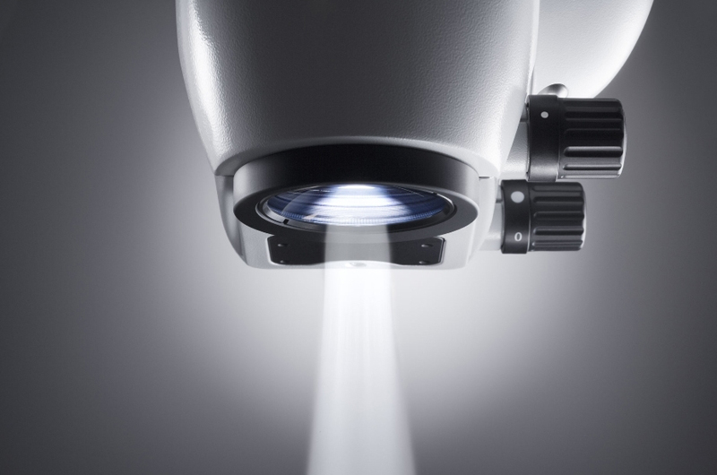
it magnifies and illuminates
allowing a perfect view of the operating field.
a better view. „
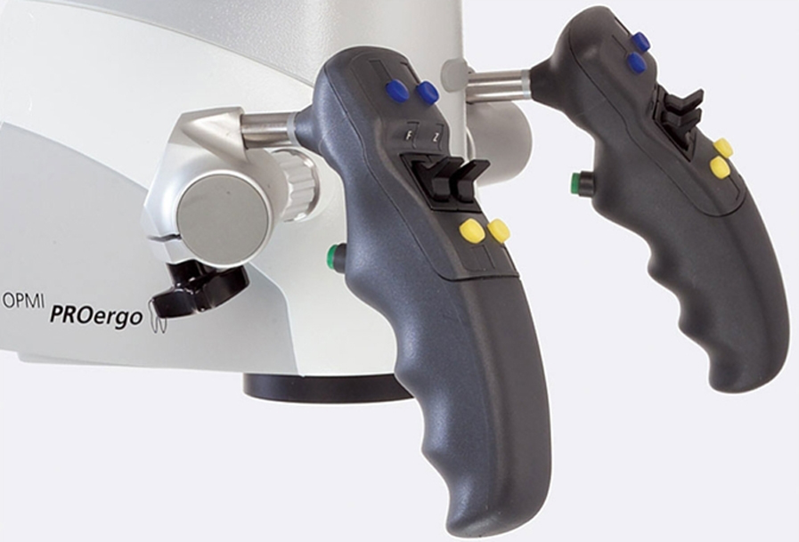
Carl Zeiss PROergo Microscope
THE ONLY WAY TO SPEED-UPYOUR DAILY ROUTINE.
From the beginning of my profession I always wondered how I could operate only with naked eye, in dentistry and oral surgery, within an operating field of few square millimetres.
I quickly turned to technology-tools: magnification devices (loupe), which enabled magnifications till 4 times with a quite good lighting.
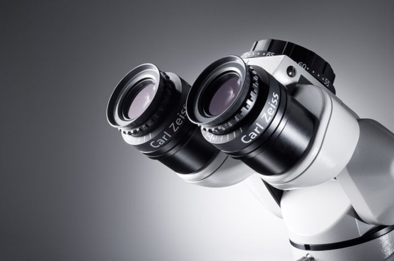
Finally in the mid '90s the crucial turning point: the stereo microscope and the real change of my operating routine. Magnification from 3 to 20 times, a refined optics and a perfectly illuminated operating field by xenon light.
Over the years I saw my technical skill holding on and at the moment my daily routine is microscope-focused.
What kind of Microscope
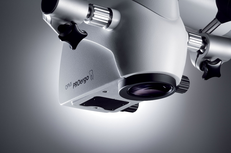
Our work doesn't take place on a flat surface (for example like in ophthalmic surgery), but inside a cavity. Therefore in microscopic dentistry and in minimally invasive surgery or oral maxillofacial surgery, it's a topic issue to get the possibility to quickly change the focal length (distance from the object), i.e. the distance from the work surface. Having the right equipment, is fundamental. Having a wide range of focal lengths is ke and an electronic zoom is recommended over a mechanical one; the larger the range the better, to permit maximum adaptability to a variety of clinical situations; I'm using a Zeiss ProERGO.
Requirements for microscoping:
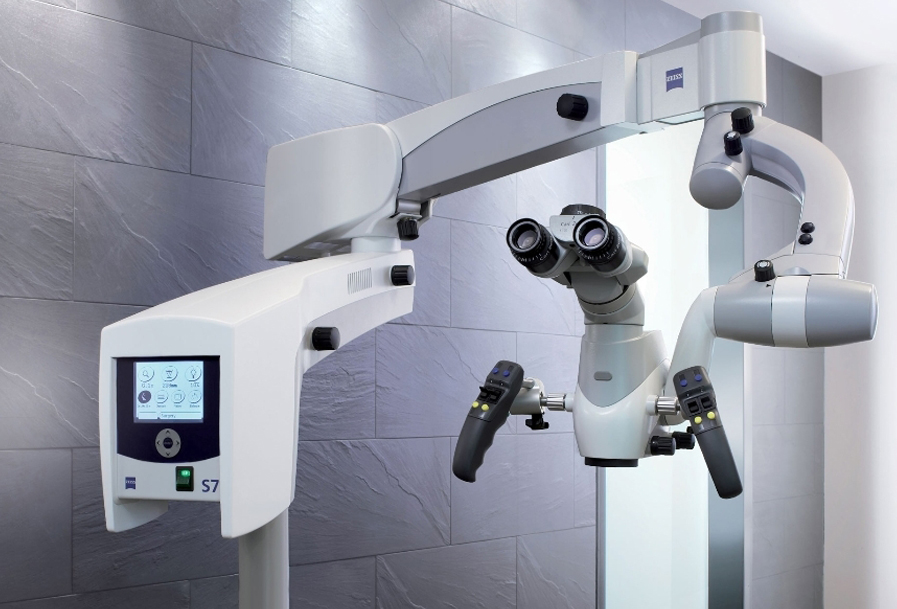
- The use of the microscope requires major changes in body positioning for the dental surgeon.
Under traditional conditions (with the naked eye or wearable optical instruments, like a helmet and magnifying lenses) the patient remains stationary and the surgeon moves around to see into the different areas of his mouth. - The conventional dentist's chair can raise and incline the patient in the Trendelenburg position (reclining with the head below the knees and waist). Instead, working with the microscope, the surgeon's body positioning is centered on the instrument (as in the solar system, with the sun at the centre): the microscope and the operator are essentially still, while the position of the patient changes depending on the area of the mouth being treated.
Therefore: the patient moves but the operator and the microscope do not! - The chair the surgeon uses allows ample head movement by permitting the head, stomach and legs to change position at the same time. There's also the possibility to add tilting, and thanks to all this flexibility the surgeon has a real time view of practically the whole mouth.
- A reflected view (like a mirror) is reserved for specific cases of microscopic dental procedures and mini-invasive surgery (the back wall of the last upper molar, an especially small mouth, etc.)
- Operating room ergonomics have developed to a point to now permit unencumbered work with the microscope. For example, to free up space, the bulky and useless rinse basin has been eliminated. Some colleagues may be surprised at this, but in fact very few patients miss it. Operating today is much more fluid and uninterrupted: rinsing has disappeared, using a dental dam is essential and suctioning is much more accurate.
- The architecture of the operating room is not unlike that of a hospital: there is ample space, furnishings are kept to a bare minimum and usable space is maximized. Portable equipment permits easy and hygienic cleaning up and, where necessary, the certainty of maintaining a sterile environment.
- Having the right operator: retinal image fusion defects create major obstacles for the operator, mainly in working without visual checks: it is better to have an eye check-up before trying your hand with a microscope!
- A training period with an expert practitioner is fundamental to shorten the steep learning curve.