


Gingival recession (receding gums) is a process in which the marginal gum tissue surrounding teeth, wears away, or pulls back, exposing more of the tooth, or of the tooth’s root. There are plenty of reasons why your gum start to recede. The problem especially affects patients who have a genetically based “fragility” of gums. Orthodontic miss-treatment takes a great part in worsening the situation, as in the following case history.

Multiple gingival recessions and compromised aesthetics
The rationale about “BLOOMING” of recessions
- Poor QUALITY of gums tissue: technically the THIN BIOTYPE. A thin, frailty gum is more likely to recede then a thick, strong one: the THICK BIOTYPE. A bad biotype is the PRIME MOVER of all recessions!
- Inadequate Brushing (link to the article): if you use an excessively stiff toothbrush, if you “harshly” brush your teeth in the wrong way (e.g. horizontally) you damage both your enamel and gums. The most affected teeth are canines and premolars. Their position on the maximum curvature of dental arch accounts for their higher vulnerability to aggressive brushing.
- Malocclusion or tooth crowding (crooked teeth): a crooked tooth always lacks of pink gingiva (the “strong” firm gum protecting teeth).
- Eating disorders (self-induced vomiting): stomach acid damages both enamel and gums.
- Alcohol, smoke, sparkling drinks, coffee, lemon or orange juice too.
- Periodontal disease: it’s a chronic inflammation disease of the “scaffold” of gums (periodontitis). It destroys bone and gum.
- Trauma due to piercing of lip, tongue, frenulum, cheek.
- Scurvy (link to the article): deficiency of vitamin C.
- Poor dental care worsens periodontal disease.
- Orthodontics : Orthodontist often doesn’t realize he is moving your tooth towards the external (buccal) part of the alveolar process. Meanwhile your tooth “out of the blue” looses its external bone support (the buccal bone plate) and gums are receding. It doesn’t happen of course immediately after orthodontic treatment. Until some years later gums of canines start disappearing. But your Orthodontist to be blamed for his bad job, already cashed in and quickly…faded away in your memory!

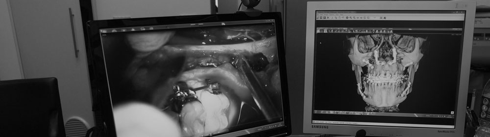
surgical planning
Types of gingival recession
“The professional point of view”
The most commonly used classification is the Miller’s one (1985). It evaluates the degree of gingival recession (considering the muco-gingival junction), the degree of underlying alveolar bone loss, the bone loss in the inter-dental area. He sorted the most common defects in 4 main CLASSES:
Class 1. Marginal tissue recession which does not extend to the muco-gingival junction with no alveolar bone loss or soft tissue loss in the inter-dental area.
Class 2. Marginal tissue recession which extends to or beyond the muco-gingival junction with no alveolar bone loss or soft tissue loss in the inter-dental area.
Class 3. Marginal tissue recession which extends to or beyond the muco-gingival junction; the inter-dental alveolar bone loss or soft tissue loss is beyond the cement-enamel junction but remains coronal to the apical extent of the marginal tissue recession.
Class 4. Marginal tissue recession which extends to or beyond the muco-gingival junction with inter-dental alveolar bone and soft tissue loss to a level corresponding to the apical extent of the marginal tissue recession.

Miller’s classification
What matters?

Miller’s classification allows Dentist to take the right treatment choice:
- complete root coverage is possible only in class 1 and class 2 recession type,
- partial root coverage is possible in class 3 type,
- NO ROOT COVERAGE is possible in class 4. type: therefore it’s ABSOLUTELY no mean to start surgery !
What’s up when gum starts to recede
Bad Aesthetic :
- tooth may appear longer than normal, because dental roots are exposed and visible;

gingival recession
- the loss of inter-dental papilla results in an awful black hole in between teeth;
- multiple recessions prevents you from a natural bright SMILE.
Functional Impairment:
- teeth become sensitive to hot and cold foods;
- brush teeth becomes a problem. The tooth neck is “naked” . Root’s gum is disappearing. Dental roots are not shielded by enamel, so they are more “sensitive” to everything!
- the patient avoids brushing aching areas. Microbial plaque amount is rocketing. The more plaque – the more inflammation, the recessions worsen.
- The final step: decay of the tooth neck.
What to do
Surgery is the ONLY way of treatment of receding gum of type I and type II, sometimes of type III, according to the Miller’s classification.
The aim of surgery: to restore the damaged “gingival frame”.
I mostly use one of the following three surgical techniques or a combination of them:
repositioning of adjacent gum tissue: I “rotate” a little tissue flap to cover the recession from a donor site to the recipient site ( by mean, the recession)
- autologous tissue graft from the roof of the mouth (palatal): I harvest a piece of palatal gum to the defect;
- combined treatments: flap+graft.
Before surgery:
- in case of severe cervical decay (link to blog) a conservative treatment (filling) is recommended;
- teach to your patient a correct brushing technique (link to blog). It is mandatory. A poor patient’s compliance will result in the failure of the best surgical results!
The Surgery
Planning of surgery grounds on the following criteria:
1. recession class according to Miller’s classification,
2. number of involved teeth,
3. bio-type of soft tissue: “the less thick the more tissue you need”
A Case History
A 42- year-old female patient,
- in good general health,
- no-smoking habit,
- showing multiple gingival recessions
- previous orthodontic treatment!
- inadequate brushing,
- dentin hypersensitivity
- thin bio-type

clinical case: multiple gingival recessions
After all previous statements, I planned a combined surgical technique:
- a Coronally Repositioned Multiple Flap with maintenance of inter-proximal papilla (Split-Full-Split)
- at the same time I’ll add a sub-epithelial connective tissue graft from the palate to the most damaged recession (on canine – 13) in order to get as much tissue as possible on this root.
The Coronally Repositioned Multiple Flap has three main characteristics:
- it does NOT require vertical releasing incision: this will NOT impair graft’s blood supply , in turn will speed-up healing time;
- the incision is planned in order to re-design the inter-dental papillae, thus restoring a nice gingival frame (the scalloping pink frame);
- split-full-split: this technique allows the sliding of an intermediate full-thickness flap including Periosteum. This provides the critical zone next to the cement-enamel junction with better vascular supply and more healing power.

Digital Flap Design

Incision are performed accordingly to the Digital Flap Design
Surgical Techniques
Once the flap’s incision is made as planned, you start the split-full-split rising of flap in three steps:
- a split thickness flap in the marginal area of crown (pink gum);
- a full-thickness flap below the muco-gingival line. Periosteum grants more mechanical support, stabilization and blood supply;
- a split-thickness flap again in the apical area, alike releasing incisions, in order to easy move up coronally the tension-free gums that passively fit into the recipient bed.
More simple:
The flap’s draw arises from the assessment of the pedicle rotation point (centre of rotation). After this you design the oblique lines of incision of papillae accordingly to the final target: to pull up – without tension – coronally the flap and cover recessions.
If you want a clear UNDERSTANDING go to the link

split-full-split flap dissection: 3 steps

split-full-split flap dissection: 3 steps

tension-free flap after periosteal release incisions
In case of severe recessions and/or a thin gingival tissue (the THIN BIO-TYPE) an autologous connective tissue graft is highly recommended in order to avoid relapse of gum recession. The number ONE best donor site to harvest connective tissue is the tuber maxillae of palate. The number TWO is the palatal gum.

Area of harvesting of connective tissue (sub-epithelial- connective graft) – connective tissue after de-epithelialization

in-layered connective tissue graft at the canine root site

sutured connective tissue graft in situ
Once the graft is properly sutured, the flap is advanced toward the clinical crowns of teeth.

final suture

healing process after 3 months

healing at the harvesting area after 3 months

before

after
Conclusions
This case required different surgeries :
- pink aesthetics re-framing,
- recessions coverage,
- a grafting procedure from the palate.
In the past patient would have been forced to a sequel of single surgical procedures, with a tooth by tooth connective tissue grafting surgery.
I performed just ONE surgery: the split-full-split graft.

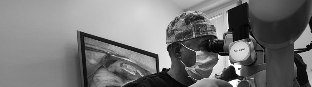
dr. Massimo Mazza
www.zeroinon.it
Prima di postare un commento leggere le Regole del Blog


